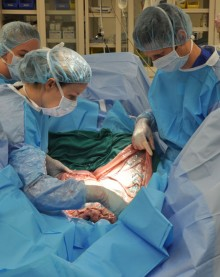
CALEC surgery, or cultivated autologous limbal epithelial cell surgery, represents a groundbreaking innovation in the treatment of severe eye damage. This pioneering procedure, performed for the first time by Ula Jurkunas at Mass Eye and Ear, utilizes stem cell therapy to restore the cornea’s surface in patients suffering from corneal injuries that were once considered untreatable. By extracting limbal epithelial cells from a healthy eye and cultivating them into a graft, CALEC surgery offers hope to those experiencing chronic pain and vision impairment caused by significant corneal defects. In clinical trials, this method has been shown to be over 90 percent effective in enhancing the quality of life for individuals with corneal injuries. With the potential to revolutionize eye damage treatment, CALEC surgery marks a key advancement in regenerative medicine and ocular health.
Cultivated autologous limbal epithelial cell treatment, popularly known as CALEC surgery, signifies a monumental step forward in ocular restoration techniques. This advanced surgical method employs stem cell therapy to mend corneal surfaces that have suffered serious injuries from various stressors, such as chemical burns or trauma. By harvesting healthy limbal epithelial cells, medical professionals can generate cellular grafts that facilitate corneal repair, thus alleviating pain and restoring vision in affected patients. The successful implementation of this treatment at renowned institutions like Mass Eye and Ear reflects a growing trend in regenerative approaches to eye care. As research progresses, the hope is to broaden the scope of this technique to assist a larger patient demographic facing similar ophthalmic challenges.
Understanding CALEC Surgery: A Breakthrough in Corneal Repair
Cultivated Autologous Limbal Epithelial Cells (CALEC) surgery represents a groundbreaking advancement in the field of corneal repair. This innovative procedure utilizes stem cells harvested from a healthy eye to repair damaged corneal surfaces, offering renewed hope for patients suffering from severe eye damage. CALEC surgery is performed at Mass Eye and Ear and involves a meticulously orchestrated process: firstly, stem cells are harvested through a biopsy from the patient’s unaffected eye; these cells are then cultured to create a cellular graft that can be transplanted into the affected cornea. This method signifies a transformative approach to treating corneal injuries that were previously deemed irreversible, particularly those induced by trauma or chemical burns.
In recent clinical trials, CALEC has shown exceptional results, with over 90% effectiveness in restoring the cornea’s surface after transplantation. For patients engaged in this trial, the outcome has led not only to improved visual acuity but also to a substantial decrease in chronic pain associated with corneal damage. The rigorous research leading to the success of CALEC surgery demonstrates a significant leap toward utilizing stem cell therapy in eye damage treatment, paving the way for potentially broader applications in ocular health.
The Role of Stem Cell Therapy in Eye Damage Treatment
Stem cell therapy has emerged as a revolutionary treatment modality in ocular medicine, particularly in addressing conditions leading to corneal damage. By leveraging the regenerative capabilities of stem cells, physicians can mitigate the effects of limbal stem cell deficiency, a condition often resulting from severe injuries or diseases. The application of stem cell therapy in these instances is particularly promising when it comes to corneal repair, as it offers a biological solution to restore the delicate architecture of the eye. Cultivated from the patient’s own tissue, stem cells help in regenerating the limbal epithelial cells that are vital for maintaining a clear and functional cornea.
At Mass Eye and Ear, significant strides in stem cell therapy have been realized through clinical trials, specifically with the CALEC procedure. The studies have highlighted how this treatment can dramatically improve the quality of life for individuals facing blinding corneal injuries by restoring not only vision but also comfort. Hence, the importance of continued research and clinical trials in this area cannot be overstated, as they hold the key to unlocking further potential treatments that can harness the power of cellular regeneration in managing severe ocular conditions.
Limbal Epithelial Cells: The Essence of Corneal Health
Limbal epithelial cells play a crucial role in maintaining corneal integrity and health. Located at the peripheral edge of the cornea, these stem cells are responsible for replenishing the surface cells that can be damaged by trauma, infections, or other ocular diseases. When these cells are depleted, as in the case of limbal stem cell deficiency, the corneal surface can develop significant complications, leading to pain, vision impairment, and even blindness. Understanding the function and importance of limbal epithelial cells is vital in appreciating the innovations brought forth by CALEC surgery at Mass Eye and Ear.
The success of stem cell therapy in repairing corneal damage relies heavily on the ability to cultivate and transplant these limbal epithelial cells effectively. The advancements in CALEC technology have showcased how these cells can be harvested and expanded to develop viable grafts that restore damaged corneas. As research continues to evolve, the integration of limbal epithelial cells into various treatments indicates a promising future for patients who suffer from ocular surface diseases, providing them with a pathway to regain their sight and improve their quality of life.
Innovations at Mass Eye and Ear: Leading the Way in Ocular Treatments
Mass Eye and Ear is at the forefront of ocular treatment innovations, especially with the application of advanced methods such as CALEC surgery. The institution has pioneered research integrating stem cell therapy to offer effective solutions for patients with severe corneal injuries that could not be remedied through traditional surgical methods. By establishing a robust clinical trial framework, Mass Eye and Ear has been able to push the boundaries of what is achievable in corneal repair, demonstrating a commitment to improving patient outcomes through cutting-edge medical practices.
The collaboration between leading ophthalmologists and researchers at Mass Eye and Ear has been instrumental in facilitating breakthroughs in eye damage treatments. The successful development and execution of the CALEC surgery underscore the importance of interdisciplinary cooperation in medical research. With continued funding from organizations such as the National Eye Institute, there lies significant potential for further advancements in human applications, ultimately expanding the horizons for patients facing vision-threatening conditions.
Prognosis and Future Directions for CALEC Surgery
The prognosis for patients undergoing CALEC surgery is promising, with clinical trials indicating high success rates in restoring corneal surfaces. As noted by researchers, the therapy has exhibited a remarkable 90% effectiveness across various stages of follow-up, suggesting a lasting impact on the participants’ vision and comfort. Such results pave the way for potential wider applications of this technique, making it a viable option for a larger patient demographic facing similar ocular challenges.
Looking ahead, further studies are essential to refine and validate the CALEC procedure. Researchers at Mass Eye and Ear emphasize the need for expansive clinical trials with diverse patient cohorts and randomized controlled design to ensure comprehensive understanding and effectiveness of the treatment. The goal is to secure federal approval to make CALEC surgery accessible at more medical centers, thereby broadening the scope of treatment options available for individuals with corneal damage.
The Importance of Clinical Trials in Advancing Eye Care
Clinical trials play a vital role in the advancement of eye care treatments, providing a structured environment to evaluate the efficacy and safety of new procedures like CALEC surgery. Through rigorous testing and monitoring, trials offer valuable insights into how innovative therapies can benefit patients, all while adhering to strict regulatory standards established by organizations such as the FDA. The recent CALEC trials reflect this commitment to careful scientific inquiry, ensuring that the benefits of stem cell therapy can be fully realized in treating corneal injuries.
Moreover, these trials facilitate collaboration among various research institutions, leading to shared knowledge and expertise that enhance the understanding of ocular diseases. By actively participating in clinical studies, leading facilities like Mass Eye and Ear contribute significantly to the foundational research needed to improve outcomes for patients suffering from severe eye conditions. Such efforts emphasize the importance of ongoing clinical evaluation in securing better treatment modalities in the realm of eye care.
Collaborative Research Efforts in Stem Cell Therapy
Collaboration between institutions is essential for the advancement of stem cell therapy in eye care, particularly as demonstrated by the partnerships formed during the CALEC surgery trials. By working with researchers from prominent organizations such as Dana-Farber Cancer Institute and Boston Children’s Hospital, Mass Eye and Ear has been able to synergize expertise and resources to innovate and optimize treatment methodologies. These joint efforts highlight how interdisciplinary approaches can significantly accelerate the development and implementation of effective therapeutic solutions.
The combined knowledge from diverse medical fields enriches the research undertaken in stem cell therapy, enabling more comprehensive studies and improved safety protocols. This collaborative approach not only enhances the scientific rigor of the trials but also fosters an environment where innovative ideas can flourish. As researchers continue to navigate the complexities of ocular stem cell treatments, interdisciplinary collaboration will be a cornerstone in achieving breakthroughs that benefit patients in need of effective eye damage treatments.
Challenges and Limitations in Limbal Stem Cell Therapy
Despite the promise shown by CALEC surgery and stem cell therapies, there are notable challenges and limitations associated with this innovative treatment. One significant hurdle is the necessity for patients to have only one functional eye from which limbal epithelial cells can be harvested. This requirement restricts the demographic of individuals who can benefit from this procedure, creating a disparity in access to this cutting-edge therapy. Additionally, the preparation time for stem cell grafts, which can take two to three weeks, may pose a waiting period that needs thorough management to ensure optimal patient outcomes.
Furthermore, while the initial results from the clinical trials have been positive, researchers recognize the need for ongoing studies to capture long-term effects and any potential complications associated with stem cell procedures. Addressing these limitations is crucial to expanding patient eligibility and refining the methodologies used in CALEC surgery. The commitment to overcoming such challenges will be critical in making limbal stem cell therapy a widely applicable and effective solution in ophthalmology.
The Future of Ocular Treatments: Stem Cells and Beyond
As advancements in stem cell technology continue to unfold, the future of ocular treatments holds immense promise. Innovations such as CALEC surgery provide a glimpse into a future where conditions once deemed untreatable can be effectively managed or even cured. The application of stem cell therapy in eye care represents not only a scientific achievement but also offers hope for those enduring the debilitating effects of corneal damage. Continuous research and successful trials will pave the way for increasingly sophisticated treatments that may soon integrate advanced technologies with regenerative medicine.
Looking beyond CALEC, researchers are actively exploring other potential applications of stem cell therapies for various ocular diseases. The field is moving towards developing allogeneic systems that could make these treatments applicable for patients with bilateral corneal damage. As the understanding of stem cell biology deepens, opportunities will arise for innovative approaches that harness the body’s own healing mechanisms. In conclusion, the future of ocular treatment guided by stem cell therapy heralds a transformative era in eye care, promising improved quality of life for countless individuals.
Frequently Asked Questions
What is CALEC surgery and how does it utilize stem cell therapy for eye damage treatment?
CALEC surgery, or cultivated autologous limbal epithelial cell surgery, is an innovative procedure that uses stem cells harvested from a healthy eye to repair damaged corneal surfaces. This treatment addresses blinding corneal injuries by creating a cellular tissue graft from limbal epithelial cells, which are critical for maintaining the cornea’s integrity. At Mass Eye and Ear, this pioneering approach has shown over 90% effectiveness in restoring corneal health, making it a groundbreaking option for patients with previously untreatable eye damage.
Who performs CALEC surgery at Mass Eye and Ear, and what is the significance of this procedure?
Dr. Ula Jurkunas, an associate director of the Cornea Service at Mass Eye and Ear, leads the team that performs CALEC surgery. This procedure is significant as it represents a new hope for individuals suffering from chronic pain and visual impairments due to corneal damage. By utilizing stem cell therapy to regenerate limbal epithelial cells, CALEC can restore the cornea’s surface and drastically improve patients’ quality of life.
What are limbal epithelial cells and why are they important in CALEC surgery?
Limbal epithelial cells are specialized cells located at the outer edge of the cornea and play a vital role in maintaining the cornea’s smooth and clear surface. In CALEC surgery, these cells are harvested from a healthy eye to create a graft for transplantation into the damaged cornea. Their regeneration is crucial, as a deficiency in these cells can lead to chronic eye damage and serious visual impairments.
What were the outcomes of the clinical trial for CALEC surgery at Mass Eye and Ear?
The clinical trial for CALEC surgery at Mass Eye and Ear yielded promising results, with complete cornea restoration achieved in 50% of participants after three months. Success rates increased to 79% and 77% at 12 and 18 months, respectively, demonstrating CALEC’s effectiveness in treating corneal damage previously deemed untreatable. Moreover, the procedure was well-tolerated, with no serious adverse events reported.
Is CALEC surgery currently available for patients, and what are the next steps for this treatment?
As of now, CALEC surgery remains experimental and is not widely available at Mass Eye and Ear or any U.S. hospitals. Future studies are necessary to expand the patient cohort and evaluate long-term outcomes before FDA approval can be pursued. Researchers hope to establish an allogeneic manufacturing process to make this treatment accessible for patients with damage to both eyes.
What role does Mass Eye and Ear play in advancing stem cell therapy for eye diseases?
Mass Eye and Ear is at the forefront of advancing stem cell therapy for eye diseases through innovative research and clinical trials, such as CALEC surgery. The collaboration among experts in ophthalmology, cell manufacturing, and research institutions like Dana-Farber ensures rigorous development of effective treatments, aiming to transition successful laboratory findings into clinical applications that improve patient outcomes.
| Key Points | Details |
|---|---|
| First CALEC surgery performed | Ula Jurkunas conducts the first CALEC surgery at Mass Eye and Ear. |
| Treatment purpose | Resolves damage from corneal injuries previously considered untreatable. |
| Clinical trial overview | The trial showed over 90% effectiveness in restoring corneal surface. |
| Mechanism of CALEC | Involves taking healthy stem cells from one eye to repair the damaged cornea. |
| Outcome statistics | Complete corneal restoration achieved in 50% of participants at 3 months, increasing to 79% and 77% at 12 and 18 months respectively. |
| Future aspirations | Plans to develop an allogeneic manufacturing process for broader applicability of CALEC treatment. |
| Current status | Still experimental; not available at Mass Eye and Ear or U.S. hospitals. |
Summary
CALEC surgery represents a groundbreaking advancement in the treatment of corneal damage, offering new hope to patients suffering from previously untreatable conditions. The results from clinical trials led by Mass Eye and Ear showcase the efficacy and safety of this innovative technique, which uses stem cells to restore the corneal surface. As researchers aim for broader applications and future studies, CALEC surgery could significantly improve many patients’ quality of life, transforming the landscape of ophthalmic treatments.
